
Utah's PC-MIL Jumps Cancer Localization from 51% to 93% Without Retraining
A new pathology MIL framework treats the spatial scale of supervision as a design axis, boosting cross-context accuracy from 77% to 96%.
 Pro
Takeaways
- PC-MIL reframes whole-slide MIL by making the spatial scale of supervision a tunable design variable
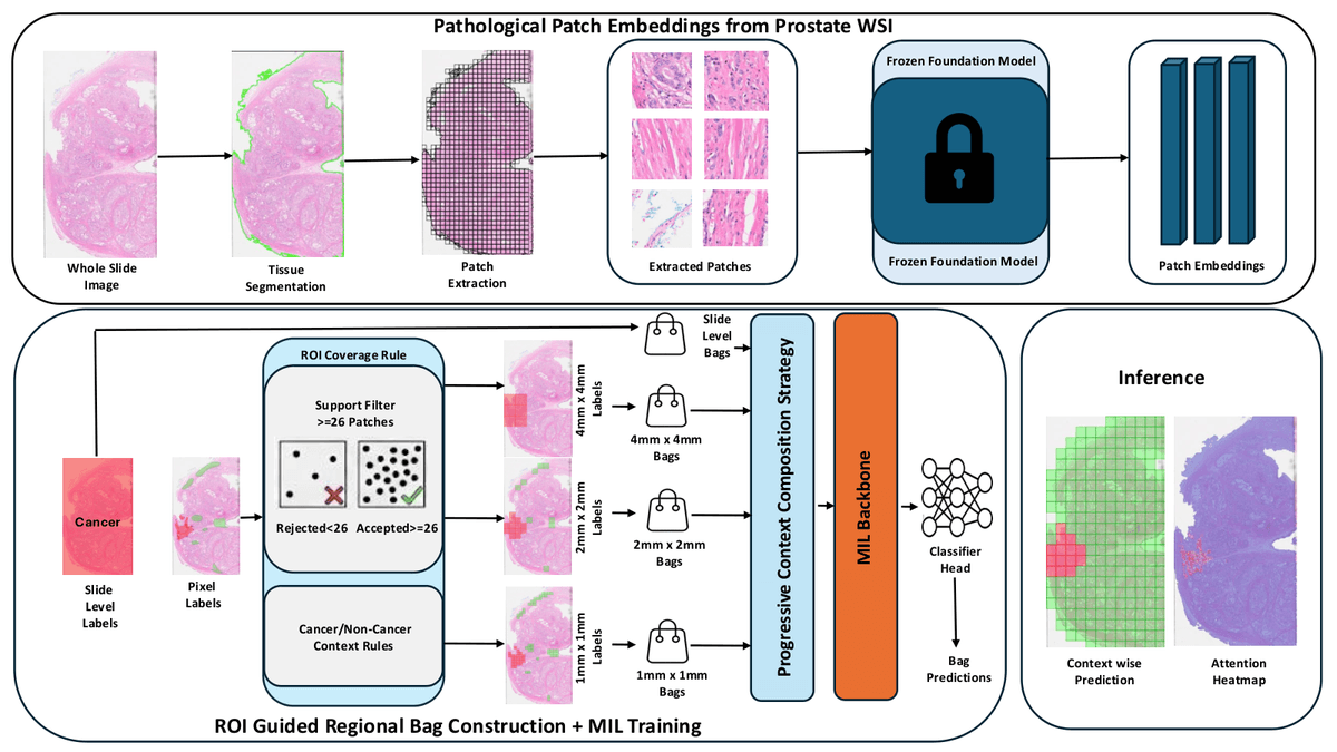
- Uses fixed 20x UNI2-h features and defines bags at 1x1, 2x2, and 4x4 mm anatomical regions
- Slide-only ABMIL gets 99.46% slide B-A but drops to 63% on 1x1 mm regions
- Balanced mix (60/24/8/8) reaches 96.16% average B-A across four evaluation scales
- Localization agreement with expert annotations jumps from 51.70% (ABMIL) to 93.18% (PC-MIL)
- Validated on 1,476 prostate WSIs from five public datasets; code not yet released
Slide-level Multiple Instance Learning (MIL) is the workhorse of computational pathology: bundle a gigapixel whole-slide image (WSI) into a bag of patches, apply a single global label, and let attention figure out what matters. It works well for slide classification, but it hides an uncomfortable truth. The model only ever learns whether cancer exists somewhere on the slide, not where, how much, or how it is arranged. A new paper from the University of Utah, Instapath, and Tulane called PC-MIL argues that this mismatch between global supervision and millimeter-scale clinical reasoning is the real bottleneck, and proposes treating supervision scale itself as a first-class training knob.
Why slide-level MIL quietly breaks down
The core issue the authors call out is identifiability. A global positive label is consistent with many spatial configurations of tumor evidence, so the learning problem is underconstrained. Attention-based aggregators like ABMIL and clustering-augmented variants like CLAM soften the rigidity of max pooling, but they still optimize a single global objective, which leaves spatial extent unconstrained by the loss.
Clinically, that is a problem. Pathologists reason about tumor burden, focal lesions, and architectural patterns inside bounded regions, and 1 to 2 mm thresholds are commonly used to define actionable surgical margins. Train a model only to answer the slide-level yes/no question and you get near-perfect slide accuracy with attention maps that spread diffusely across the tissue rather than concentrating on tumor-dense regions.