
University of Utah's AC-MIL Grades Cardiac MRI Scans Like a Radiologist Would
A weakly supervised framework cracks open the multiple instance learning black box, telling clinicians exactly why a cardiac MRI scan failed quality checks.
 Pro
Takeaways
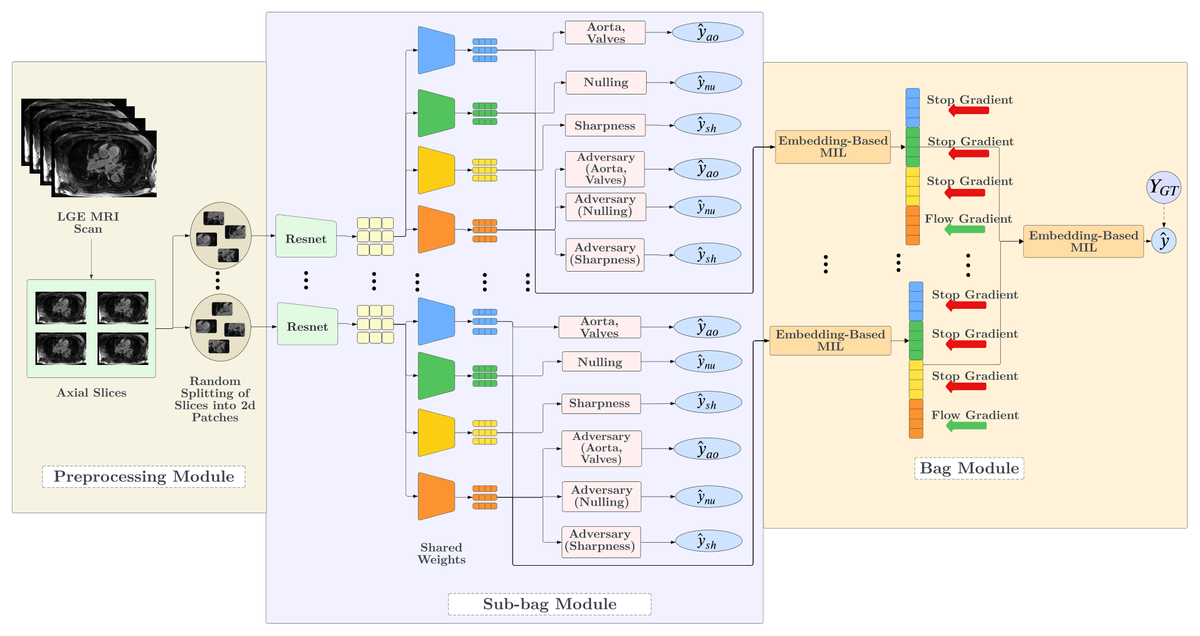
- AC-MIL decomposes cardiac MRI quality scores into clinically defined concepts using only volume-level labels (paper).
- Three predefined concepts: Sharpness, Myocardium Nulling, Aorta and Valve Enhancement, plus an unsupervised residual branch.
- Adversarial gradient reversal purges known concepts from the residual, preventing information leakage.
- Spatial Attention Diversity loss forces each concept head to attend to distinct anatomical regions.
- Achieves 0.66 QWK on 775 LGE-MRI scans, competitive with prior HAMIL-QA at 0.63.
- Code to be released on acceptance; framework is domain-agnostic for weakly supervised medical imaging tasks.
Automating medical image quality control sounds boring until you realize it directly affects whether patients get the right treatment. A team from the University of Utah just published AC-MIL, a framework that grades cardiac MRI scans and explains its reasoning in the same terms a radiologist would use, all while training on nothing but a single quality score per volume.
Atrial fibrillation is the most common form of cardiac arrhythmia, affecting an estimated 3 to 5 million people in the U.S., with projections suggesting the number could surpass 12 million by 2030. Late Gadolinium Enhancement (LGE) MRI is the imaging workhorse used to detect the fibrotic tissue that drives the disease and to plan catheter ablation procedures. Diagnostic utility depends heavily on image quality. Patient movement and inconsistent breathing introduce motion artifacts, while suboptimal pulse sequence parameters and inadequate contrast administration can further degrade clarity. Manual evaluation is labor-intensive and hard to fit into a fast-paced clinical workflow.
Where existing MIL models break down
Multiple Instance Learning (MIL) has become the default weakly supervised approach for this problem: split a 3D volume into patches, learn attention weights over them, and pool everything into a single vector that predicts a volume-level label. It works, but current state-of-the-art methods map localized visual evidence to a single opaque global feature vector. That black-box output fails to provide actionable feedback on specific failure modes, obscuring whether a scan degrades due to motion blur, inadequate contrast, or missing anatomical context.