
UIC Turns Unstable Vision Transformers Into Medical Classifiers With 29-Point Accuracy Gain
A curriculum over network depth improves fine-grained anatomical classification on limited medical data, with transformer models gaining more than 16 accuracy points.
 Pro
Takeaways
- Progressive growing trains block-structured networks in phases, activating deeper layers only after shallow ones converge.
- ViT-B/32 accuracy on SOS staging jumped from 0.3624 to 0.6542 with the same architecture and loss.
- ResNet-101 improved from 0.8465 to 0.8600 while using 93.2% of the baseline compute budget.
- Method validated on 723 expert-annotated CBCT scans across five fusion stages.
- Generalization confirmed on CIFAR-10, where ViT-B/16 gained 3 points with the same schedule.
- No code or checkpoint released; training-dynamics change only, no architecture or loss modifications.
Teaching a neural network to read a skull scan the way an orthodontist does is harder than it sounds. A new paper from the University of Illinois Chicago and the University of Orléans tackles a specific clinical bottleneck, staging the fusion of the spheno-occipital synchondrosis (SOS), and shows that how you train a model matters more than which model you pick. Their progressive growing recipe turns wobbly Vision Transformers into stable classifiers and squeezes extra accuracy out of ResNets, all without touching the architecture or the loss function.
The bone that tells time
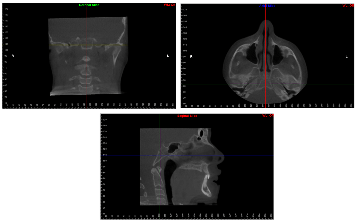
The spheno-occipital synchondrosis is a cartilage joint in the cranial base that fuses gradually through adolescence. Because its closure tracks pubertal growth, orthodontists use it to time appliances, orthognathic surgery, and growth modification, especially when other maturity indicators are outside the CBCT field of view. Prior clinical work built expert-annotated datasets of 723 CBCT scans labeled by two orthodontists and an oral and maxillofacial radiologist to enable automation.
Fusion is a continuum rather than a set of discrete states, which is where the trouble starts. Radiologists integrate weak cues like endocranial fusion, trabecular continuity, and residual scar patterns across scales, and adjacent stages look almost identical. That makes SOS staging a textbook fine-grained visual recognition problem, and one where standard deep networks tend to overfit or converge unstably on the transitional stages that clinicians care about most.