
Koblenz Researchers Beat U-Net Limits, Hitting 0.914 Dice With 10 Patients
A U-Net trained with TotalSegmentator organ masks pushes AAA segmentation Dice from 0.64 to 0.91 without changing the architecture.
 Pro
Takeaways
- Anatomy-aware U-Net for AAA segmentation lifts Dice from 0.637 to 0.914 without architectural changes (paper).
- Uses TotalSegmentator to build organ exclusion masks that gate the training loss.
- Loss is only computed on anatomically plausible pixels, removing implausible regions rather than penalizing them.
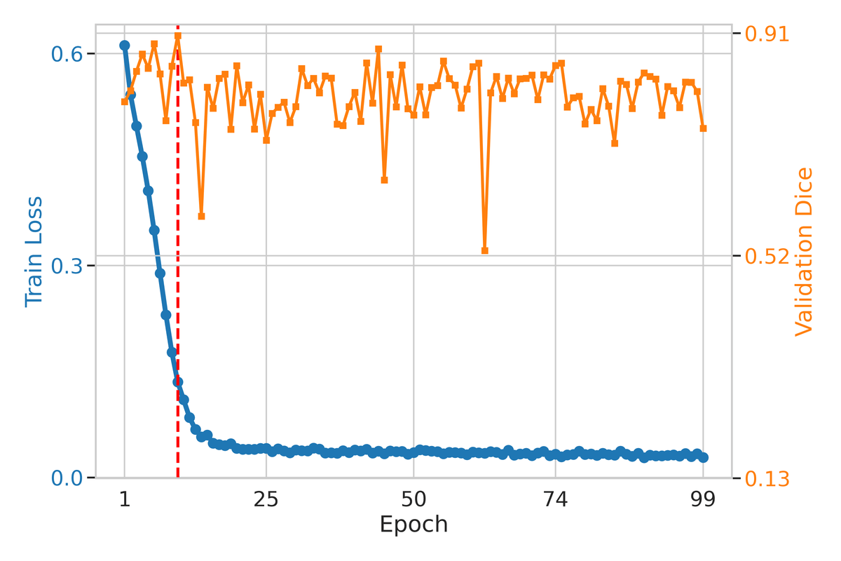
- Trained on just 10 patients yet cuts slice-wise Dice standard deviation from 0.442 to 0.033.
- Largest gains occur near bowel, vertebrae, and kidneys where thrombus intensity mimics organ tissue.
- Includes 3D mesh reconstruction and VMTK centerline analysis for clinical morphological metrics.
Segmenting abdominal aortic aneurysms (AAAs) on CT angiography looks like a solved problem on paper. In practice, tiny datasets, low-contrast vessel walls, and organs that share the same intensity range as vascular tissue still trip up standard U-Nets. A new paper from the University of Applied Sciences Koblenz proposes a cheap fix: tell the network which parts of the abdomen it is not allowed to predict on, using free anatomical labels from TotalSegmentator.
The approach takes a plain 2D U-Net from 0.637 mean Dice to 0.914 on the test set, with almost no engineering overhead and only 10 training patients. No architectural tricks, no extra manual labels, no attention modules.
Why AAA segmentation keeps breaking
An abdominal aortic aneurysm is a dilation of the aorta with a rupture mortality rate exceeding 80%, so accurate delineation of the outer wall matters for surgical planning. Segmentation performance degrades badly near anatomically similar structures such as vertebrae, bowel, and kidneys, producing false positives. Thrombus, the clotted material inside the aneurysm, has intensity values close to muscle and bowel content, so a network trained only on image intensity happily lights up nearby organs.
Prior work tried to fix this with multi-organ co-segmentation, shape priors, or attention gates, but those approaches often increase annotation effort, model complexity, or computational cost. That is a bad trade when you only have 20 annotated patients.