
Duke's FullTilt Detects Proteins 1000x Faster by Skipping 3D Reconstruction
A new open-set detection framework skips the 3D tomogram entirely, running zero-shot particle picking on raw tilt-series hundreds of times faster.
 Pro
Takeaways
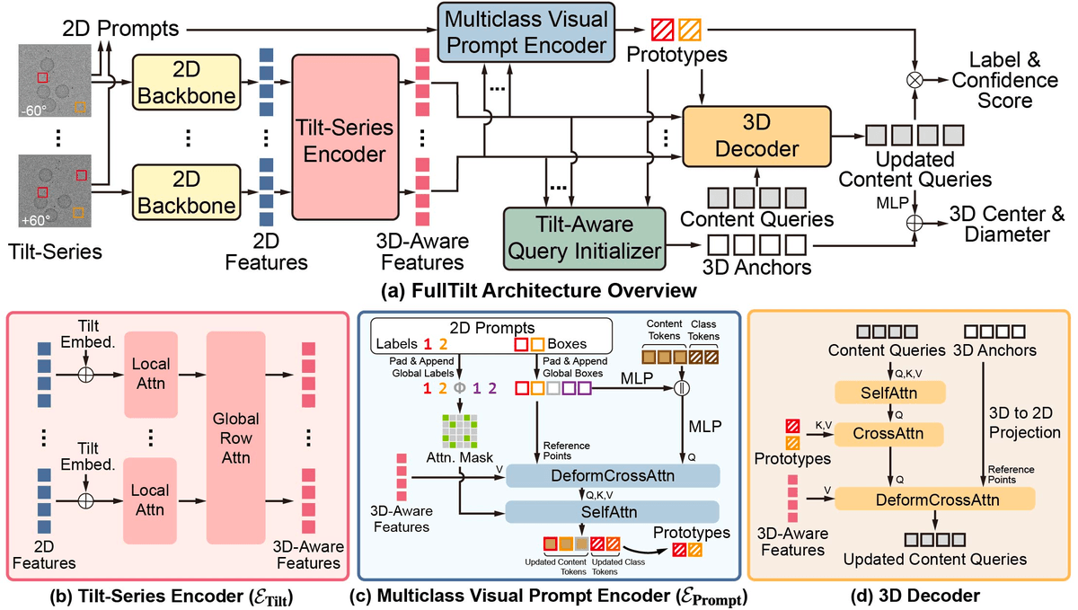
- FullTilt runs open-set cryo-ET particle detection directly on 2D tilt-series, skipping 3D tomogram reconstruction entirely.
- Inference drops from thousands of seconds to under one second, using roughly 6x less VRAM than baselines.
- A DETR-style architecture with four new modules: tilt-series encoder, multiclass prompt encoder, tilt-aware query init, geometric primitives.
- Matches or beats TomoTwin, ProPicker, and CryoSAM on CZII, EMPIAR-10304, and EMPIAR-10499.
- Ablations show cross-tilt fusion is essential: without the tilt-series encoder, detection performance collapses to zero.
- Limitations include weak performance on very small or non-globular particles; code will be released publicly.
Cryo-electron tomography (cryo-ET) has become one of structural biology's most powerful tools for imaging proteins in their native cellular environment, but pulling individual molecules out of the noisy 3D volumes it produces is painfully slow. A new framework called FullTilt, from Duke University, tackles that bottleneck by discarding a long-standing assumption in the field: that you need to reconstruct a 3D tomogram before you can detect anything inside it.
FullTilt runs detection directly on the raw 2D tilt-series, and the payoff is dramatic. The framework achieves inference speeds hundreds to thousands of times faster than current state-of-the-art open-set macromolecule detection methods, while matching or beating them on accuracy across three real-world datasets.

Why current cryo-ET pipelines crawl
To understand the leap, it helps to know how cryo-ET data is produced. The specimen is physically rotated inside the microscope during acquisition, and a set of 2D projections from multiple angles is collected, called a tilt-series. The tilt-series is then computationally aligned and reconstructed to yield a 3D volume, or tomogram. Downstream tools then hunt through that volume for copies of specific macromolecules (called particles) and average them together to get high-resolution structures.
Traditional detectors like DeepFinder or crYOLO are closed-set: every new protein target requires fresh annotations and a retrained model. To escape that treadmill, recent methods such as TomoTwin, ProPicker, and CryoSAM adopted an open-set approach where you supply a visual prompt (a reference subvolume) and the model finds matches without retraining. The problem is scale. A tomogram is enormous, and strict VRAM constraints prohibit loading an entire volume into memory, so current methods rely on sliding-window inference, which balloons runtime. Others apply SAM to 2D slices along each orthogonal axis, but they still exhibit computational complexity that scales with the tomogram's volumetric dimensions.