Google Research Maps 166,000 Neurons in a Fruit Fly Brain
Google Research and HHMI Janelia released a wiring diagram of 166,000 neurons and 125 million synapses spanning the male fruit fly brain and nerve cord.
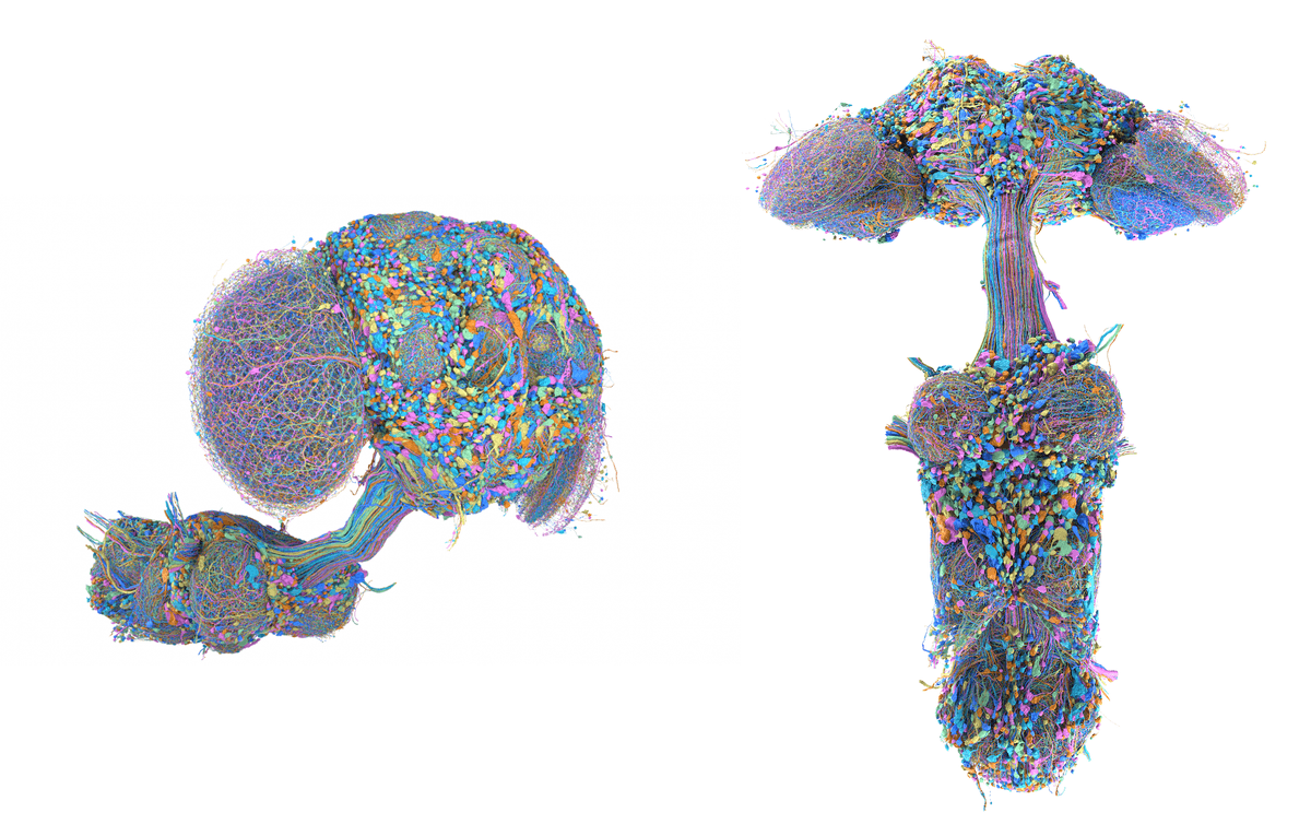
- Google Research and HHMI Janelia published the complete male fruit fly connectome: 166,000 neurons, 125 million synapses.
- Largest cellular brain map ever, covering brain, optic lobes, and ventral nerve cord (spinal cord analog).
- Built with flood-filling networks and the new PATHFINDER system trained partly on synthetic neurons.
- Dataset is freely available and browsable in open-source Neuroglancer.
- Enables direct male/female comparison for courtship, aggression, and sexually dimorphic circuits.
- Same pipeline is now mapping elephantnose fish and larval zebrafish, the first vertebrate whole-brain targets.
A decade of imaging, AI-driven segmentation, and painstaking human proofreading just produced the largest cellular-scale brain map ever built. Google Research, in partnership with HHMI Janelia, has released a complete connectome of the male fruit fly, covering the brain and the ventral nerve cord that connects it to the body.
With over 166,000 neurons and 125 million synaptic connections, this is the largest brain map by neuron count to date. The work is published in Cell as Sexual dimorphism in the complete connectome of the Drosophila male central nervous system, and the dataset is freely browsable.
What a connectome actually is
A connectome is a wiring diagram: every neuron drawn in 3D and every synaptic contact between them cataloged. Building one requires sectioning a brain into millions of thin slices, imaging each section, and stitching the images together. From those electron microscope stacks, algorithms must trace each individual neuron through the volume without confusing it with its neighbors, then identify synapses at the pixel level.
This map includes the ventral nerve cord, the structure analogous to the spinal cord, which lets researchers trace a signal from an eye or antenna all the way to the muscles that make the fly walk, sing, or fight. Prior fly connectomes stopped at the brain boundary, leaving that sensory-to-motor path incomplete.

How the reconstruction pipeline works
The core approach uses flood-filling networks: convolutional neural networks that start at a single pixel and identify every other pixel belonging to the same object. The technique has been refined over several years, with the current system called PATHFINDER. A recent addition incorporated synthetic neurons into training data, improving speed and accuracy without requiring more hand-labeled ground truth. Training on procedurally generated neuron shapes helps the model handle rare morphologies it rarely encounters in real tissue.
Progress in neuron count tells the story concisely:
- 2019: fully automated reconstruction of a female fruit fly brain released
- 2020: human-verified map of half a female brain, 25,000 neurons and 21 million connections
- 2025: 166,000 neurons across the full male CNS, roughly a 6x increase over the half-brain map
The output is served through Neuroglancer, Google's open-source WebGL viewer for petabyte-scale volumetric data. Raw meshes, synapse tables, and cell-type annotations are all available from the Janelia dataset page.
Why the male brain matters when a female map already exists
A female fly connectome already exists via the FlyWire project. Having both sexes mapped enables comparative work that a single connectome cannot support. Where circuits differ between sexes, researchers can now examine the structural basis of behaviors like courtship and aggression. Where circuits look identical, the two datasets give the first honest read on how much individual variation exists between two nominally equivalent brains.

The neuron AOTU008, shown above, illustrates this directly. The male version has two additional projections that are invisible without accurate 3D reconstruction.
What researchers can do with it
Three companion papers landed alongside the connectome, applying the map to visual systems, taste, and social behavior. Broader use cases now within reach:
- Tracing a stimulus from sensory neurons through interneurons to motor output, with no guesswork about intermediate wiring
- Building biologically constrained neural network models whose architecture is copied from the fly rather than hand-designed
- Comparing circuit motifs across species as vertebrate maps come online
- Grounding pharmacology and behavioral experiments in a known wiring substrate
From insects to vertebrates
The same pipeline is now being aimed at animals with nervous systems closer to ours. A Columbia-led study in Nature used equivalent tooling to map part of the elephantnose fish hindbrain, combining the static connectome with activity recordings to produce the most complete mechanistic model of learning in a vertebrate brain yet published. Larval zebrafish are the next target: their brains are small enough to map end-to-end with current techniques, and their larval-stage transparency allows neural activity measurements during experiments, captured in the ZAPBench dataset.
What changes from here
Two assumptions worth revising. Connectomes were long treated as too expensive for routine science, requiring years of human effort just to verify and annotate neural shapes. Synthetic training data and improved auto-proofreading are cutting that cost fast enough to put vertebrate whole-brain maps on the table. Static wiring diagrams were also sometimes dismissed as insufficient for understanding behavior; the fish study shows they compose cleanly with activity recordings to yield mechanistic learning models.
The human brain, at 86 billion neurons, sits roughly six orders of magnitude beyond what is currently tractable. The jump from a 25,000-neuron hemibrain to a 166,000-neuron full CNS took about five years, and the tools that made it possible are open source and pointed at larger targets.
Comments